

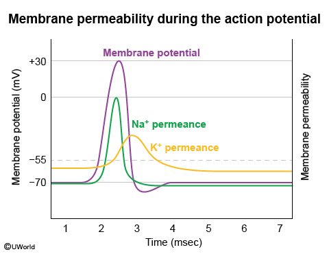
The graph depicts the potential voltage changes across a cell membrane; these changes (depolarization, repolarization, hyperpolarization, and resting potential) are collectively known as the action potential. The action potential occurs due to changes in the membrane permeability to Na+ and K+ ions. The membrane potential of an excitable cell (eg, nerve and muscle) cycles through the following stages:
Resting potential (Choice A): Usually equal to -70 mV. It is maintained by high resting membrane permeability to K and low permeability to Na. K efflux occurs via non-gated K channels (leak channels). While at the resting potential, the inner side of the membrane is negatively charged with respect to the outer surface of the membrane.
Depolarization: Occurs due to opening of voltage-gated Na channels with rapid influx of Na into the cell. The large influx of Na leads to an increased positive charge inside the membrane known as depolarization (Choice B). Overshoot refers to the maximal value of the action potential during which the membrane potential obtains a positive value (approximately +35 mV) (Choice C).
Repolarization (Choice D): Results from closure of Na channels and simultaneous opening of K channels. This causes a sharp decrease in the membrane permeability to Na and a significant increase in K permeance that exceeds that of the resting membrane. K efflux is responsible for returning the membrane potential back to the resting potential.
Hyperpolarization (Choice E): Occurs because the voltage-gated K channels remain open for a short time after repolarization is completed. The membrane potential thus becomes more negative than the normal resting potential and approaches the K equilibrium potential of -85 mV. When the voltage-gated K channels close, the membrane potential returns to the resting value maintained by the non-gated K channels.